

Introduction
1.Toxoplasma gondii 是猫科动物的肠道球虫,因其滋养体形似弓形而得名。
2.其分类地位是:Kingdom Protista>Subkingdom Protozoa>Phylum Apicomplexa>Class Sporozoa>Order ?>Family Toxoplasmatidae> Toxoplasma gondii
3.该虫呈世界性分布,可感染鸟类、鱼类、哺乳动物和人,引起Toxoplasmosis.
4.人群平均感染率33%,有5亿人抗体阳性。我室血清学检查数据显示重庆地区孕妇抗体阳性率20%以上。
Morphology
Trophozoite (tachyzoites and bradyzoites)

Indirect fluorescent antibody test. Slide shows endozoites exposed to antibody containing serum and then treated with fluorescein conjugated anti-human globulin. There is a zone of light green fluorescence around each organism. The cytoplasm appears reddish as it was counterstained with Evan's blue. ×1000. Enlarged by 5.4.

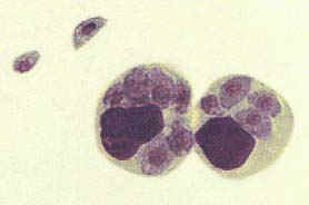
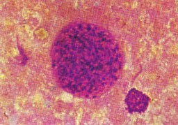
In the centre is a macrophage filled with endozoites. This stage is also known as the pseudocyst. Giemsa stain. ×1000. Enlarged by 9.6.
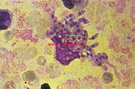
A ruptured macrophage which has liberated endozoites. Many are still lying close to the macrophage nucleus. Giemsa stain. ×1000. Enlarged by 9.6.

A ruptured macrophage with the endozoites dispersed in the background. Some parasites are undergoing division. The large dense body is the macrophage nucleus. Giemsa stain. ×1000. Enlarged by 5.4.

A macrophage containing endozoites and some free endozoites, stained with acridine orange. The cytoplasm of the endozoites consisting mostly of RNA takes up a red color and the nucleus consisting mainly of DNA takes up a yellowish green color. ×1000. Enlarged by 5.4.
Intracellular tachyzoites of Toxoplasma gondii.
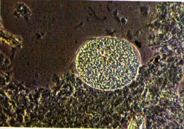
Oocyst


Freshly passed Toxoplasma oocysts. Most of the oocysts have a single spororblast. Interference contrast. ×400. Enlarged by 5.4.
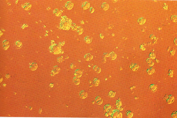
Toxoplasma oocysts after incubation in vitro for 12 hours. Most of the oocysts have now sporulated. Interference contrast. ×400. Enlarged by 23.4.
cyst
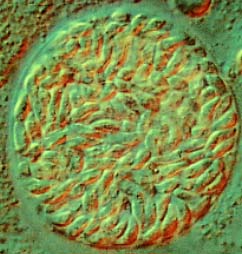
Toxoplasma cyst from mouse brain. The cyst has partly detached from the brain tissue. Phase contrast. ×400. Enlarged by 5.4.

Toxoplasma cyst from mouse brain. The cyst has completely detached from the brain tissue. Phase contrast. ×400. Enlarged by 5.4.

Toxoplasma cyst from mouse brain. Showing the argyllophilic nature of the cyst wall after silver staining. This is the best method for demonstration the cyst wall. ×1000. Enlarged 5.4.

Toxoplasma cyst from mouse brain. Showing the PAS positive cystozoites. The cyst wall is faintly visible. This is the best method for demonstrating the cystozoites. ×1000. Enlarged by 5.4.
Toxoplasma cyst from mouse brain after H and e stain. The cyst wall is faintly visible and the nuclei appear as dark bodies inside the cyst. ×1000. Enlarged by 5.4.